
Micrographs from those working in Soil Micromorphology will be dispalyed on this page.
It is hoped that this will be a regular feature and perhaps
something which will promote discussion. Contributions for future
months are welcome.
[for details -see below]
The first micrograph from Paul Goldberg was displayed for the
first time on 27th November 1997. This is a test to check that
the system works, - in particular to assess whether there are
difficulties encountered by those overseas because of the size of
the image.
All the images will be displayed in miniature form on this page.
Please click on the miniature to display the full image. It
should also be possible for you to download the image in the
normal way.
December 1997 [800k]
- click on miniature to reveal full size image
![[hard to describe]](gifs/du93min2.jpg)
Micrograph supplied by Dr Paul Goldberg: Department of
Archaeology, Boston University, 675 Commonwealth Ave, Boston,
MA2215, USA: Email: paulberg@bu.edu
The micrograph shows reworked hearth material (charcoal,
burned clay soil pellets and some ash) from Dust Cave, Alabama.
This is a Palaeoindian through Archaic site in Northern Alabama,
excavated by Dr. Boyce N. Driskell, University of Alabama,
Tuscaloosa.
This is quite
a large image and may take a little time to load. It is displayed
in full 16.7 million colours. If problems of loading arise, it
will be possible to reduce the size by about 40% by reducing the
number of colours to 256. Please inform geo.micro@uea.ac.uk, and we
shall attempt to put an alternative format.
![]()
January 1998 [257k]
- click on miniature to reveal full size image
 Micrograph supplied by Drs Keith Tovey
and David Dent: School of Environmental Sciences, University of
East Anglia, NORWICH, NR4 7TJ, UK:
Email: k.tovey
@uea.ac.uk
Micrographs shows open cellular fabric of kaolinite particles
forming under Avicennia africana mangrove under
hypersaline conditions at Bitang Bolon, The Gambia

 If you have stereo coloured specs then
you may wish to look at the stereo version to gain a full
impression of the openness of the fabric,
![]()
February 1998 [70k]
- click on miniature to reveal full size image
 Micrograph supplied by George Macleod [Email:
g.w.mcleod@stir.ac.uk]
at University of Stirling as part of a research project by Prof.
Donald Davidson. The micrograph taken in plain polarised light
shows Enchytraeid excrement infilling of a root chanel.
March 1998 [275k]
- click on miniature to reveal full size image
![]() Micrograph supplied by Louis Bresson [Email:
bresson@jouy.inra.fr]
It represents a high resolution bulk density image obtained from
and X-radiograph of impregnated soil. A special procedure is used
for calibration.
Micrograph supplied by Louis Bresson [Email:
bresson@jouy.inra.fr]
It represents a high resolution bulk density image obtained from
and X-radiograph of impregnated soil. A special procedure is used
for calibration.
April 1998 [335k]
- click on miniature to reveal full size image
 Micrograph supplied by Sacha Mooney [Peat
Technology Centre, Faculty of Agricultural Engineering,
University College, Dublin,
,Email: sjmooney@iveagh.ucd.ie]
A thin section of milled peat from West Boora, County Offaly,
Ireland comprising of amorphous grains, plant/root cross sections
and moss fragments
May 1998 [256k]
- click on miniature to reveal full size image
 Micrograph supplied by Tristam Hardman
[School of Environmental Sciences, University of East Anglia,
Norwich, UK] Email: t.hardman@uea.ac.uk]
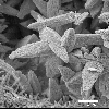
Microfabric of Halimeda
SEM images of the formation of HMC cements within Halimeda
utricles
June 1998 [256k]
- click on miniature to reveal full size image
 Micrograph supplied by Keith Tovey [School
of Environmental Sciences, University of East Anglia, Norwich,
UK] Email: k.tovey@uea.ac.uk]
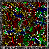
The micrograph shows effect of image analysis on a sample of
consolidated kaolin. The analysis is automatic and delineates
regions of similar orientation.
We wish to encourage people to submit examples of their micrographs for display as future Micrographs of the Month.
The preferred method is to submit the micrograph in digital
form attached to an Email to geo.micro@uea.ac.uk
![]()
This page is maintained by the Geotechnical Micromorphology and
Microanalysis Centre. geo.micro@uea.ac.uk
![]()